[最も人気のある!] human eyeball dissection eyeball cut in half 104117
27/02/19 · In a number of ways, the human eye works much like a digital camera Light is focused primarily by the cornea — the clear front surface of the eye, which acts like a camera lens The iris of the eye functions like the diaphragm of a camera, controlling the amount of light reaching the back of the eye by automatically adjusting the size of the pupil (aperture)Dissecting an Eye Before dissection, allow the students to a have a good look at the Eye and see if they can identify any parts of the Eye Before commencing any kind of dissection on animal material, always read and implement any Health & Safety measures Ensure all equipment and work surfaces are cleaned carefully and thoroughly after use Before making the fi rst incision, cutPopular because of the similarity to the human eye Ages 12 Find More Eye dissection guide $100 As low as $075 Sheep's Eye Dissection Inside & Out PowerPoint PPT Presentation of each structure Create a side view diagram of a human eye with appropriate labels The cow eye dissection is a great lesson to use as part of your unit on the nervous Guide – Vision (pdf), See
Chicago Il Sliced Human Bodies Cow Eyeball Dissection
Human eyeball dissection eyeball cut in half
Human eyeball dissection eyeball cut in half-2' Sheath of the optic nerve paasiog into the sclerotic;4 Ciliary muscle, its radiating portion ;


Cow S Eye Dissection Step 4
Well this is an interesting question As a person who has some experience cutting eyeballs I would say it would depend on the 1 Sharpness of the knife 2 Thickness of the knife 3 The eyeball stability 4 Pressure inside the eyeball The answers t18/09/17 · Flip the anterior half of the eye over so that the front of it is facing upward Using a pair of sharp scissors, cut the cornea from the eye along the boundary where the cornea meets the sclera When the scissors have cut in far enough, a clear fluid will start to seep out – this is the aqueous humorAnterior part of the human eye, with label of posterior chamber at right Schematic diagram of the human eye (posterior chamber labeled at upper left) Details;
05/03/14 · In the video, Exton walks through a dissection of a horse's eye for the At Bristol science center, showing off everything from the flopping flap of the iris to the flaky retina cellsTest both halves of your mind in this human anatomy quiz Immediately beneath the sclera is an underlying vascular layer, called the uvea, that supplies nutrients to many parts of the eye One component of the uvea is the ciliary body, a muscular structure located behind the iris that alters the shape of the lens during focusing and produces the aqueous humour that bathes the anteriorAnterior View Posterior View Name _____ Dissection Internal Anatomy – Day 2 Procedure 1 Using a sharp scalpel, cut through the sclera around the middle of the eye so that one half will have the anterior features of the eye and the other half will contain the posterior (see figure 2) The inside of the eye cavity is filled with liquid This is the
Human eye models are widely used in teaching of Human Anatomy in schools and colleges Eye models and charts are also used to teach optometrists and opticians and also for patient education All eye models are of medical qualityColor in the diagram as you learn what parts make up an eye Article from08/04/21 · Eyeball (Bulbus oculi) The eye is a highly specialized sensory organ located within the bony orbitThe main function of the eye is to detect the visual stimuli (photoreception) and to convey the gathered information to the brain via the optic nerve (CN II)In the brain, the information from the eye is processed and ultimately translated into an image
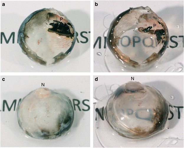


Optical Clearing Of The Eye Using The See Deep Brain Technique Eye


Cow Eye Dissection Guide
Do not remove the object stuck in eye;Avoid giving aspirin, ibuprofen or other nonsteroidal, antiinflammatory drugs These drugs thinSheep Eye Dissection The anatomy of the human eye can be better shown and understood by the actual dissection of an eye One eye of choice for dissection, that closely resembles the human eye, is that of the sheep Sheep eyes are removed at the time the animal is slaughtered and then preserved for later use Differences between the two eye types will be mentioned as the dissection


Cow Eye Dissection Cows Compared To Humans Without Moving Your Head Look Up Look Down Look All Around Six Muscles Attached To Your Eyeball Move Ppt Download


Eyeballdissection Redwoods81
18/09/17 · Eye Anatomy The human eye is one of the the most complex and Benjamin Franklin had two pair of glasses, one for near and one for far He got tired of changing them, so he cut the lenses in half and repositioned them so that he could see both near and far using the same glasses – the first bifocals!External Cow Eye Dissection DAY 1 MOO?When we had finally finished cutting the eyeball in half a mysterious marbleshaped object spilled out It was called the lens When you look through the lens the object on the other side is seen upside down This is because our brains correct the image so that we see it the correct way up This information is delivered to the rain through the optical nerve The optical is located at the back


Chicago Il Sliced Human Bodies Cow Eyeball Dissection


Activities And Answer Keys Ck 12 Foundation
The human eye is remarkable Although it is small in size, the eye arguably provides us with the most important of the five senses – vision Vision occurs when light enters the eye through the pupil With help from other important structures in the eye, like the iris and cornea, the appropriate amount of light is directed towards the lens Just like a lens in a camera sends a message toThe anatomy of the human eye can be better shown and understood by the actual dissection of an eye One eye of choice for dissection, that closely resembles the human eye, is that of the sheep Differences between the two eye types will be mentioned as the dissection is completed Begin the dissection by gathering the equipment and supplies listed here Materials Needed (sheep eyeColor in the diagram as you learn what parts make up an eye Jul 26, 16 Our eyes are one of the most important parts of our body they allow us to see!
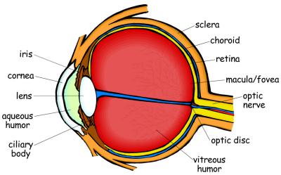


How Vision Works Eye Science Projects Experiments Hst


Pin On Anatomy Medical Contains Graphic Photos
Do not rub or apply pressure to eye ;Chicago, Illinois Sliced Human Bodies, Cow Eyeball Dissection Halfinchthick crosssections of a man and woman hang in transparent panes, like deli slices of Olive Loaf It's education, Chicago style!Crosssection illustration of an eyeball showing the eye cut in half with retina, vitreous, lens Image size Clear Select the amount of time you plan to use the image Usage length 0 $ Eyeball crosssection anatomy illustration #AN0005a quantity Add to cart Add to LightBox SKU N/A Categories Antsegment anatomy, Eye Anatomy Illustrations, Postsegment anatomy s anatomy
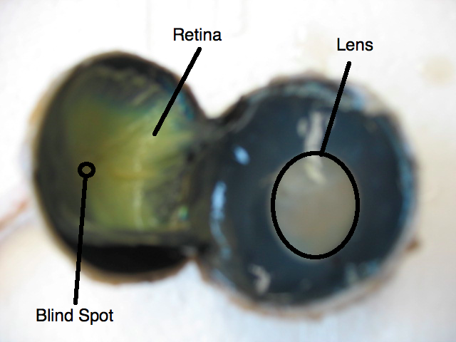


Sheep Eye Disection Mohtadi Alkhaliq
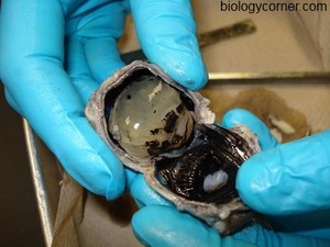


Cow Eye Dissection
The anatomy of the horse, a dissection guide Horses Fig 32 View of the Lower Half of the Eight Adult Human Eye, divided horizoktallt THROUGH THE MIDDLE MAGNIFIED FOUR TJMES {A, TUomSOtl) 1 The cornea;Horses Anatomy DISSECTION OF THE EYEBALL 265 sclerotic by being colourless and transparent The choroid coat will be recognised as the black layer lying subjacent to the sclerotic It does Fig 36 View of the Lower Half of the Right Adult Human Eye, divided horizontally THROUGH THECut Out Do not include these words Filter eyeball Stock Photos and Images 57,757 matches Page of 578 An image of a blue eye ball Eye icon close up view of eyeball Human eye anatomy, detailed illustration Isolated on a white bacground eye macro Anatomy of the eye, cross section and view of fundus Detailed illustration eyeball with shadow on white background vision, eyeball


Eyedissection Soccer007avery


Cow Eye Dissection
These muscles allow the eye to move in different directions so that the animal can see more of its surroundings without turning its head 3 Trim the fat and muscle from around the eye Be careful not to cut the optic nerve on the back of the eye 4 Using scissors or a scalpel, carefully cut the eye in half Separate the front from the back of the eye a1' Its conjunctiTal layer ;Anatomy Eye from Front;
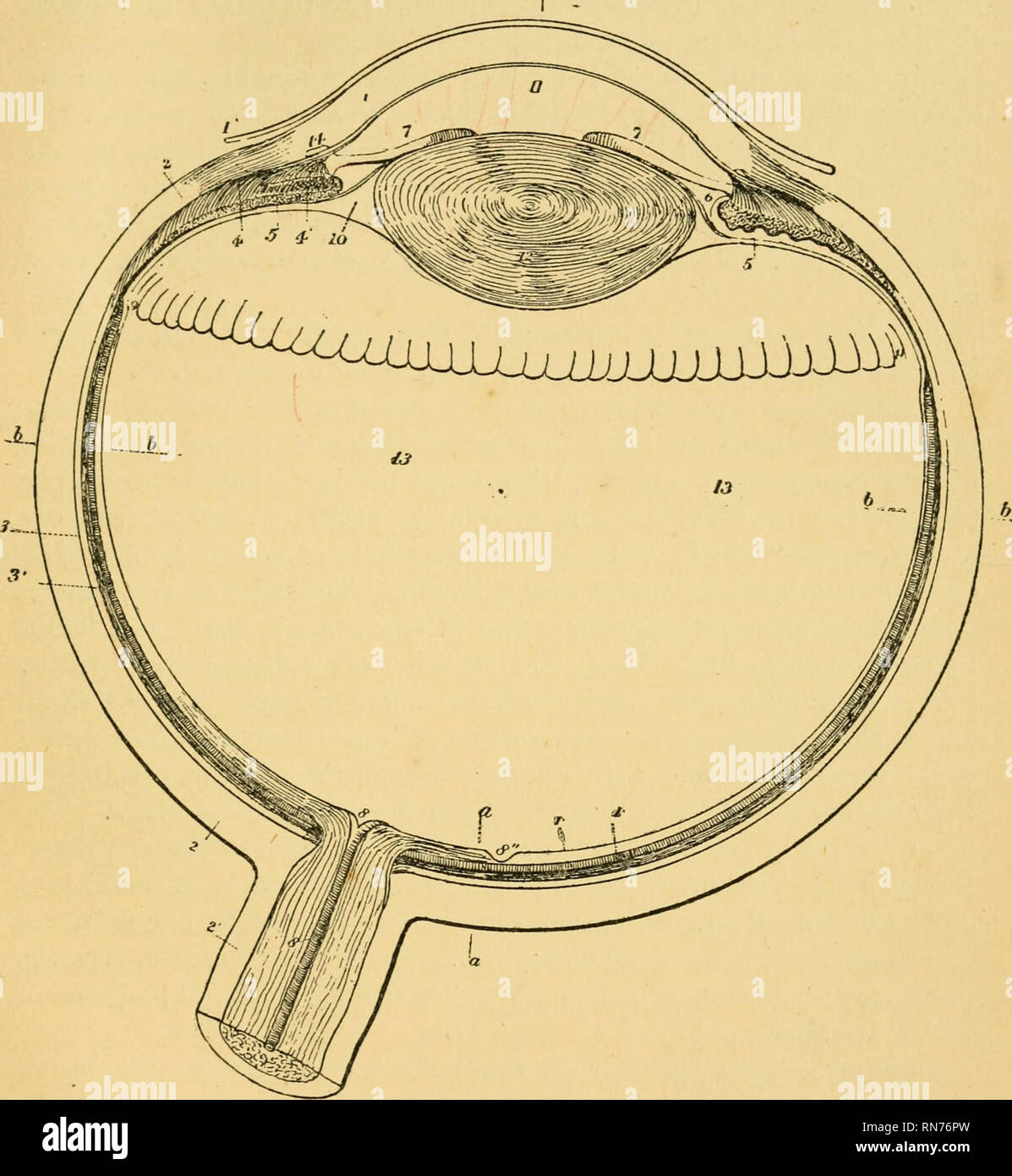


The Anatomy Of The Horse A Dissection Guide Horses Horses Anatomy Dissection Of The Eyeball 265 Sclerotic By Being Colourless And Transparent The Choroid Coat Will Be Recognised As


10lbu9x2hsjorm
Museum of Science and Industry Address 57th St, Chicago, IL Directions Museum of Science and Industry 57th St and Lake Shore Dr Phone Admission Adults $182 Place the eyeball on the dissecting tray and gently hold it with two fingers at the cornea and the optic nerve Cut a crosssection through the sclera slightly behind the middle of the eyeball so that the eyeball is separated into an anterior half and a posterior half Only cut through the wall of the eyeball, not through the entire globeUse a pencil to roll it into a halfcylinder Color the central vessels red ) Glue tabs 21) Glue optic nerve to back of sclera 22) Glue muscle to top of sclera You can color the muscle pinkishred if you want to, but leave the ends white 23) Glue eyeball to background piece NOTE Look at it from the side, making sure that the background piece is making


Human Eye Definition Structure Function Britannica


Lab A Sheeps Eye Purpose To Explore The
3 3' The choroid;With the advance of technology, visiontesting equipment hasColor in the diagram as you learn what parts make up an eye Jul 26, 16 Our eyes are one of the most important parts of our body they allow us to see!


Sheep Eye Dissection Procedures Pdf Free Download


Dissection Middle School Science Blog
Latin Camera posterior bulbi oculi T8 A A T 6794 FMA Anatomical terminology edit on Wikidata The posterior chamber is a narrow space behind the peripheral part of the iris,Brain and Eyeball Dissection Lab Report Gabrielle Warner Introduction Why are the sheep brain and cow eye used to study anatomy?08/10/11 · This is clearly shown in the corrosion cast of a cut face of the human choroid in Figure 21a (Zhang, 1974) The corresponding venous lobules drain into the venules and veins that run anterior towards the equator of the eyeball to enter the vortex veins (Fig 21b) One or two vortex veins drain each of the 4 quadrants of the eyeball The vortex veins penetrate the sclera and


Cow Eye Dissection


Pdf Dissection Of A Mouse Eye For A Whole Mount Of The Retinal Pigment Epithelium
Cow Eye Dissection May 13 1 Cow Eye Dissection Outreach Activity I Overview In this activity, students use a cow eye to learn anatomy of the eye Students dissect the cow eye and learn the different structures that make up the human eye versus the cow eye Throughout this activity students also learn how light travels through the eye into the visual system After the dissection,Download this stock image The anatomy of the horse a dissection guide Horses;While the cow's mammalian eye is a close parallel to the human eye, it does have some slight differences Through dissection and examination, students should recognize that the cow's pupil, for example, is oval, while the human pupil is round They should also notice that cow's irises are always brown, while human irises come in many colors, including blue, brown and green If


Anatomical Histological And Computed Tomography Comparisons Of The Eye And Adnexa Of Crab Eating Fox Cerdocyon Thous To Domestic Dogs


Eyedissection Soccer007avery
29/01/19 · First, we need to cut the clear plastic part of the googly eye and then attach it to the iris area with glue Then we will make Ciliary artery and, for that, we have to paint half of the foam ball using (Bright red) acrylic color For making Optic nerve we will use toilet paper roll First, we will cut in half and we need to cut it half into horizontally then we will make a roll and attach itLearn how to dissect a cow's eye in your classroom This resource includes a stepbystep, hints and tips, a cow eye primer, and a glossary of terms The Exploratorium is more than a museum Explore our online resources for learning at home At the Exploratorium, we dissect cows' eyes to show people how an eye works This Web site shows photos and videos of a dissection If youANATOMY Eye in Cross Section Click on a label to display the definition Tap on the image or pinch out and pinch in to resize the image ) Home;


Eye Dissection Ppt Download


What Do People Who Are Blind See
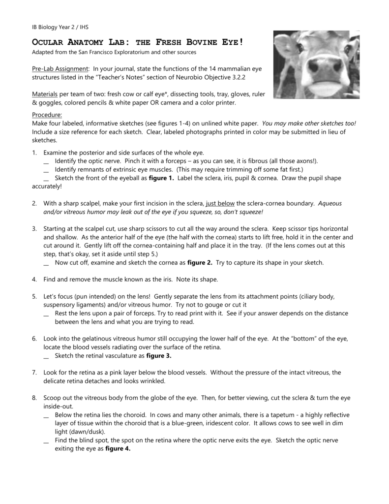
Gently place a shield over the eye to protect it Cut away the bottom part of a paper cup and tape this piece to the area around the eye Wear this to protect your eye until you get medical help do not rinse with water;COW'S EYE dissection page 6 Now take a look at the rest of the eye If the vitreous humor is still in the eyeball, empty it out On the inside of the back half of the eyeball, you can see some blood vessels that are part of a thin fleshy film That film is the retina Before you cut the eyeCOW EYE DISSECTION 1 Examine the outside of the eye You should be able to find the sclera, or the whites of the eye This tough, outer covering of the eyeball has fat and muscle attached to it 2 Locate the covering over the front of the eye, the cornea When the cow was alive, the cornea was clear In your cow's eye, the cornea may be cloudy or blue in color 2 Cut away the fat and


If I Cut An Eyeball Will It Burst Or Will It Cut Neatly In Half Like A Ball Of Jelly Quora


Cureus The Eye Of Horus The Connection Between Art Medicine And Mythology In Ancient Egypt
Cow eyes are typical dissection specimens used in lab to study eye anatomy because they are structurally and functionally similar to human eyes Students explore the external and internal anatomy, learning how structures work together to create images from incoming light A preserved cow eye dissection can be carried out in 1–2 class periods and only requires basic dissectingHuman Eye model, 5 times life size with 8 parts Removable parts of the anatomical human eye model include Upper half of the sclera with cornea and eye muscle attachments Both halves of choroid with iris and retina Eye moreImage of a human eye showing the blood vessels of the bulbar conjunctiva Hyperaemia of the superficial bulbar conjunctiva blood vessels The conjunctiva is a tissue that lines the inside of the eyelids and covers the sclera (the white of the eye) It is composed of unkeratinized, stratified squamous epithelium with goblet cells, and stratified columnar epithelium The conjunctiva is


Cow Eye Dissection


Salmonids In The Classroom Salmon Dissection
Place eye in dissection tray Examine the eye and identify the following parts The sclera – the whites of the eye, the tough, outer covering of the eyeball Fat and muscle surrounding the eye The cornea – the covering over the front of the eye The iris, the coloured part of the eye The pupil, the dark oval in the middle of the iris07/02/ · Each completed game level gives 1 knowledge point in Human eye anatomy The maximum number of points (7 knowledge points) is achieved when you pass all 7 levels You'll get a bronze medal when you complete a level 2 times and a silver medal after 5 completed rounds A gold medal will be received after 10 completed rounds In Human eye anatomy, the maximumBy using the sheep brain and cow eye, learning becomes easier I am a hands on learner and when i can touch or see things i am able to make connections


Cow S Eye Dissection Instructions


Mammal Eye Dissection Human Eye Eye
Eye c Use your scissors to cut around the middle of the eye, cutting the eye in half You'll end up with two halves On the front half will be the cornea The cornea is made of pretty tough stuff—it helps protect your eye It also helps you see by bending the light that comes into your eye d Once you have removed the cornea, place it on


The Beauty Of The Human Eye Iris Structure Similar Planet Easy To Share


Dissecting A Cow S Eye


Sheep Head Dissection Brains Tongues And Eyes Middle School Science Blog


Eyeballdissection Beanie17
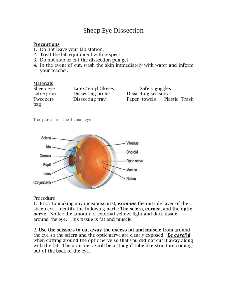


Sheep Eye Dissection


Cataract Surgery Wikipedia


Dissected Human Eye Page 4 Line 17qq Com
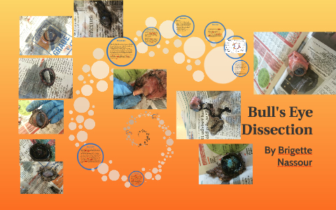


Bull S Eye Dissection By Brigette Nassour


Human Eye Definition Structure Function Britannica


Cow Eye
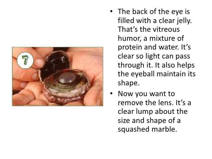


Cow S Eye Dissection Instructions


The Human Heart 10x808 Thingscutinhalfporn


Cow S Eye Dissection Step 4
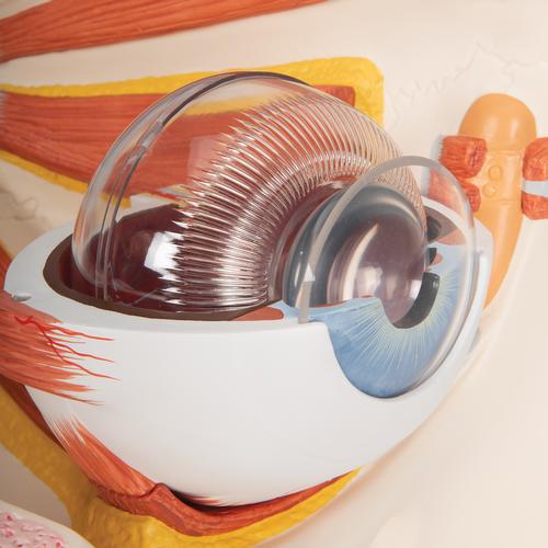


Anatomical Teaching Models Plastic Human Eye Models Eye With Optic Nerve Model


Eye Paper Dissection Scienstructable 3d Dissection Model Digital Lesson Life Science Science Projects Biology


Human Eye Cut In Half Page 6 Line 17qq Com


Cow S Eye Dissection Step 7


Human Eye Definition Structure Function Britannica


Eye Dissection Ingridscience Ca
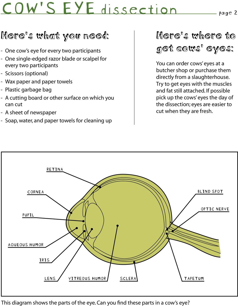


Cow S Eye Dissection Dissecting A Cow S Eye Step By Step Instructions Safety First Pdf Free Download


Anatomy Of The Human Optic Nerve Structure And Function Intechopen


Eyedissection Soccer007avery
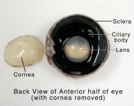


Cow Eye Dissection Anatomy Project Hst Learning Center


Real Human Eye Dissection Page 1 Line 17qq Com
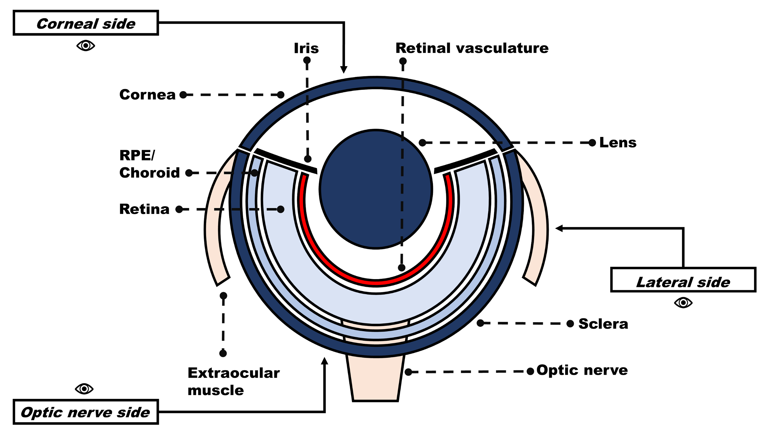


Studying Diabetes Through The Eyes Of A Fish Microdissection Visualization And Analysis Of The Adult Tg Fli Egfp Zebrafish Retinal Vasculature Protocol


Eye Anatomy Glaucoma Research Foundation


Acp Best Practice No 164 Journal Of Clinical Pathology


Cow Eye Dissection Youtube


Dissection Of Harbour Seal Lenses A Lens Lying On The Vitreous Download Scientific Diagram
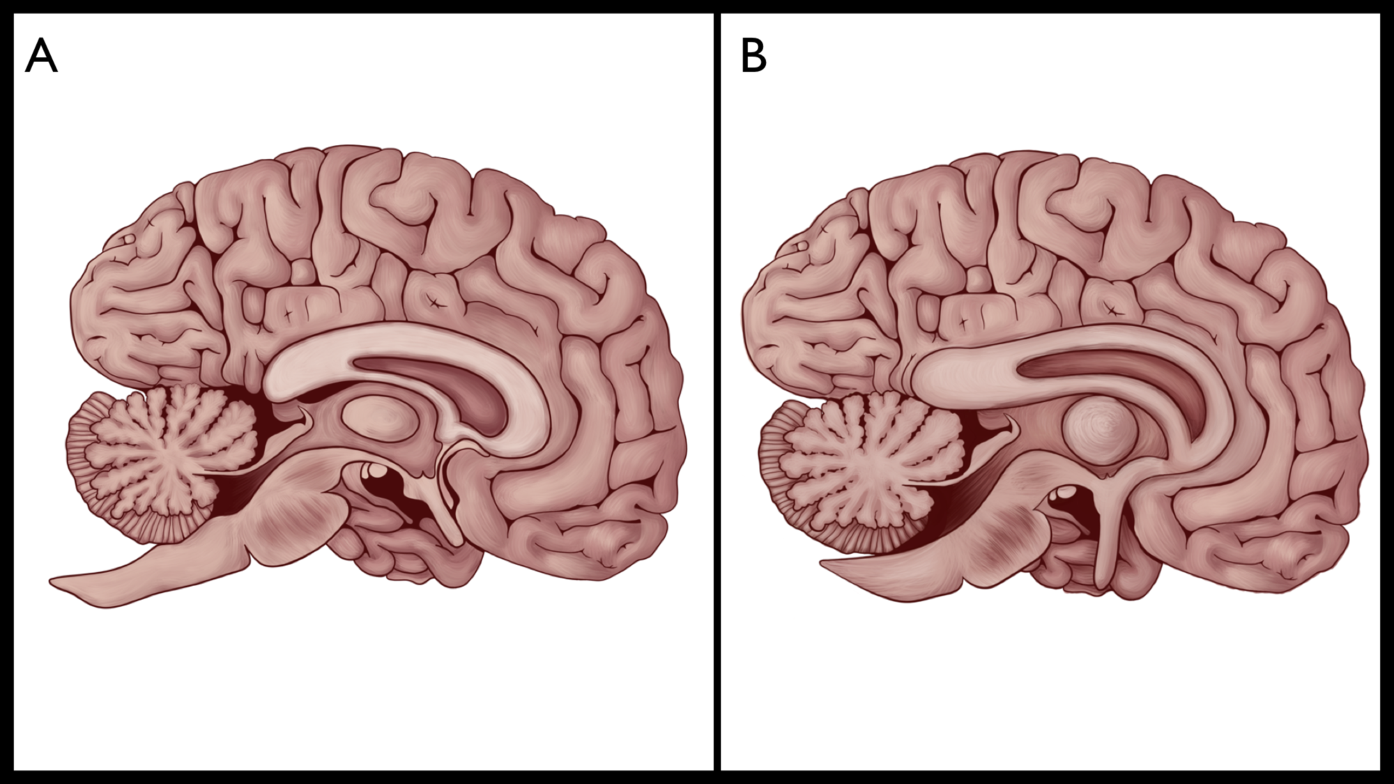


Cureus The Eye Of Horus The Connection Between Art Medicine And Mythology In Ancient Egypt


Activity 2 4 1 Exploring The Anatomy Of The Eye


How Forensic Scientists Once Tried To See A Dead Person S Last Sight Smart News Smithsonian Magazine


Cow Eye Dissection Student Cut 2 For Lesson Plan Youtube


Cow Eye Dissection Anatomy Project Hst Learning Center


Axis Scientific 5x Enlarged Human Eye Model Eyeball Anatomy Multi Part Structure Dissects To 7 Anatomically Correct Parts Of Eye Includes Removable Stand Detailed Product Manual 3 Year Warranty Amazon Com Industrial Scientific


Cow S Eye Dissection Instructions


Eye Structure And Function In Cats Cat Owners Merck Veterinary Manual


10lbu9x2hsjorm
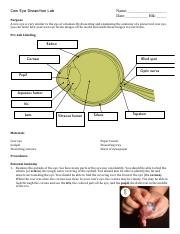


Cow Eye Dissection Lab Cow Eye Dissection Lab Name Date Blk Purpose A Cow Eye Is Very Similar To The Eye Of A Human By Dissecting And Examining The Course Hero
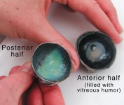


Cow Eye Dissection Anatomy Project Hst Learning Center


Real Human Eye Dissection Page 3 Line 17qq Com


Human Eye Cut In Half Page 3 Line 17qq Com


Real Human Eye Dissection Page 1 Line 17qq Com
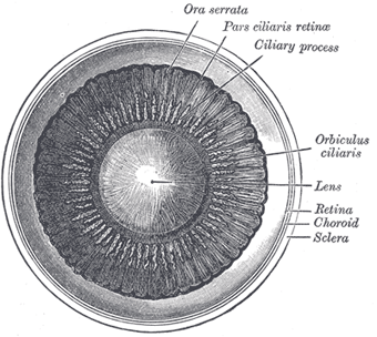


The Tunics Of The Eye Human Anatomy


Cow Eye Dissection Youtube


Sheep Eye Disection Mohtadi Alkhaliq


Activities And Answer Keys Ck 12 Foundation
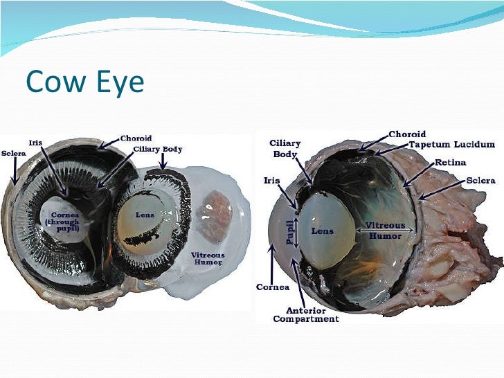


19 Best Cow Eye Dissection Labeled


Cow Eye Dissection Kit For Kids Animal Anatomy Labs Hst


Cow S Eye Dissection Step 5


What S Inside An Eyeball Eyeball Dissection We The Curious Youtube


コメント
コメントを投稿